


Digestive System of Frog Diagram & Functions for NEET Students
The concept of the Digestive System of Frog is essential in biology and helps explain real-world biological processes and exam-level questions effectively.
Understanding Digestive System of Frog
Digestive System of Frog Diagram refers to the complete arrangement of organs and structures that help break down food, absorb nutrients, and remove wastes in frogs. This concept is important in areas like amphibian digestion, animal physiology diagrams, and NEET comparative biology questions.
Digestive System of Frog: Parts and Mechanism
The digestive system of frogs is well developed and helps in the ingestion, digestion, absorption, assimilation, and egestion of food. Frogs are carnivorous animals that mainly feed on insects, worms, snails, and other small organisms. Their digestive system includes a complete alimentary canal along with associated digestive glands that help in breaking down food into simpler absorbable substances.
The different parts of the digestive system of frog and their functions are explained below.
Alimentary Canal of Frog
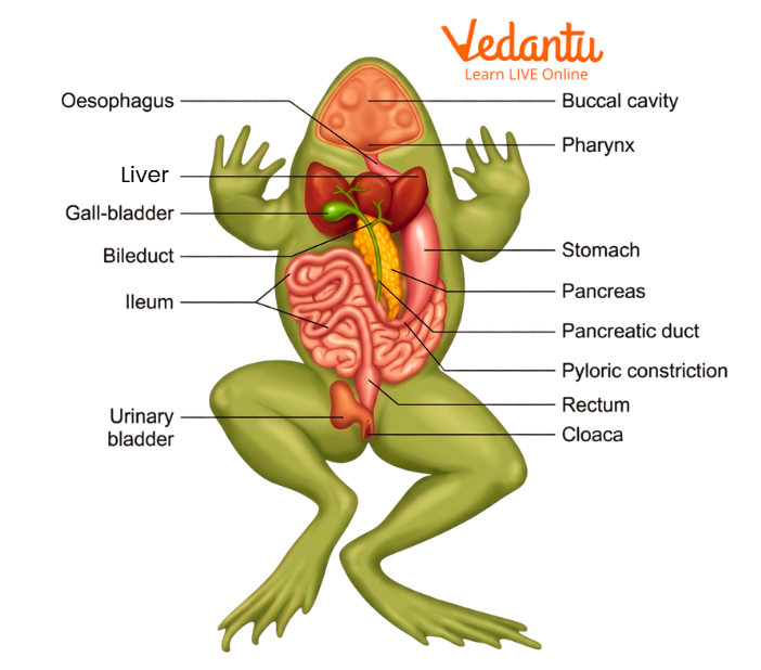
In frogs, the alimentary canal is complete. It is a long, coiled tube that extends from the mouth to the cloaca. It includes the following parts:
Mouth
Buccal cavity
Pharynx
Oesophagus
Stomach
Small intestine
Large intestine
Cloaca
Each part performs a specific role in the digestion process.
Mouth
The alimentary canal begins with the mouth, which is a wide opening extending across the snout. It is bounded by two jaws. The upper jaw is fixed, while the lower jaw is movable, allowing the mouth to open and close during feeding.
This broad mouth helps the frog capture prey quickly.
Buccal Cavity
The mouth opens into a wide, shallow buccal cavity lined with ciliated columnar epithelium. This lining contains mucous glands that secrete mucus to lubricate food. Frogs do not have salivary glands, so no saliva is produced in the buccal cavity.
The buccal cavity mainly helps in holding prey and passing it backward into the digestive tract.
Teeth
Frogs do not have teeth in the lower jaw. Teeth are present only in the upper jaw on the maxillae and premaxillae, and there are also two patches of vomerine teeth on the roof of the buccal cavity.
Features of Frog Teeth:
Small, pointed, and backwardly directed
Similar in shape, so they are homodont
Fixed to the jaw bone, not set in sockets
Used for holding prey, not chewing
These teeth prevent captured prey from escaping. Frogs are also polyphyodont, which means their teeth are replaced many times during life.
Internal Nostrils
The roof of the buccal cavity contains two openings called internal nostrils or internal nares. These connect the buccal cavity to the nasal chambers and allow air to pass during respiration.
Tongue
The tongue of frog is large, muscular, sticky, and protrusible. It is attached at the front of the lower jaw, while its posterior end remains free and bifid.
Functions of the Frog Tongue:
Captures prey with its sticky surface
Can be rapidly protruded and withdrawn
Helps in swallowing food
The upper surface of the tongue contains taste buds and mucous glands. These glands make the tongue sticky, but they do not produce digestive enzymes.
Orbit Bulging
Behind the vomerine teeth, the roof of the buccal cavity shows two bulges formed by the eyeballs. During swallowing, the eyes are pushed downward into the buccal cavity. This helps press the food backward into the pharynx.
This is a unique mechanical adaptation in frog digestion.
Pharynx
The buccal cavity narrows behind into the pharynx, which opens into the oesophagus through the gullet. Together, the buccal cavity and pharynx are often called the buccopharyngeal cavity.
Important structures present in the pharynx include:
Openings of Eustachian tubes on either side, which connect to the middle ear
Glottis, a median slit behind the tongue that opens into the lungs
In male frogs, openings of the vocal sacs, which help in croaking
The glottis remains open during breathing but closes during swallowing.
Oesophagus
The oesophagus is a short, wide, and muscular tube connecting the pharynx to the stomach. It is highly distensible because its inner lining forms longitudinal folds. This allows it to expand when swallowing large prey.
Its main function is to conduct food into the stomach.
Stomach
The stomach lies on the left side of the body cavity and is attached to the dorsal body wall by the mesogaster. It is a curved, broad tube divided into two regions:
Cardiac stomach – broad anterior part
Pyloric stomach – narrow posterior part
The inner lining of the stomach forms folds that allow expansion. The stomach contains gastric glands that secrete digestive substances.
Secretions of the Stomach:
Pepsinogen from gastric glands
Hydrochloric acid from oxyntic cells
Hydrochloric acid converts pepsinogen into active pepsin, which begins protein digestion. At the pyloric end, a muscular sphincter regulates the release of food into the small intestine.
Intestine
The stomach opens into the intestine, a long and coiled tube attached to the dorsal body wall by the mesentery. It is divided into two parts:
Small intestine
Large intestine
Small Intestine
The small intestine is the main site of digestion and absorption. It is divided into:
1. Duodenum
The first part of the small intestine forms a U-shaped loop. It receives:
Bile juice from the liver
Pancreatic juice from the pancreas
These juices enter through a common hepatopancreatic duct.
2. Ileum
The ileum is long and highly coiled. It is the region where most digestion and absorption take place.
The inner lining of the small intestine contains:
Goblet cells, which secrete mucus
Absorbing cells, which absorb digested nutrients
Unlike higher vertebrates, true villi and crypts are absent, though the surface is folded to increase absorption.
Large Intestine
The large intestine or rectum is short and wide. It receives undigested food from the ileum and stores it temporarily as faeces before elimination.
Its lining contains low longitudinal folds.
Cloaca
The cloaca is a small sac-like chamber at the end of the alimentary canal. It receives:
Faecal matter from the rectum
Urinary and reproductive discharges
It opens outside through the cloacal aperture at the posterior end of the body.
Physiology of Digestion in Frog
The physiology of digestion in frog includes the following major processes:
Ingestion
Digestion
Absorption
Assimilation
Egestion
Food of Frogs
Frogs are carnivorous animals. Their diet mainly includes:
Insects
Earthworms
Spiders
Snails
Small fish
Small frogs
They capture prey with the help of their protrusible sticky tongue and swallow it whole.
Food Ingestion
When a frog spots prey nearby, it opens its mouth and quickly throws out its sticky tongue. The prey sticks to the tongue and is pulled back into the buccal cavity.
Once the prey enters the mouth:
The inwardly directed maxillary and vomerine teeth prevent escape
The pharyngeal walls contract and push the food backward
Peristaltic movements of the oesophagus move the food into the stomach
This method allows frogs to feed rapidly on moving prey.
Food Digestion
Food digestion in frogs involves both physical and chemical changes.
1. Physical digestion
Physical changes occur due to:
Peristaltic movements of the alimentary canal
Churning movements in the stomach
Pressure from the eyeballs during swallowing
2. Chemical digestion
Chemical digestion is carried out by enzymes, which convert complex food materials into simple soluble substances.
Different enzymes act on different food components:
Proteolytic enzymes digest proteins
Lipolytic enzymes digest fats
Diastatic enzymes digest carbohydrates
3. Buccal Digestion
No actual digestion occurs in the buccal cavity because:
There are no salivary glands
No digestive enzymes are secreted there
Only mucus is added, which softens and lubricates the food for easy passage.
4. Gastric Digestion
In the stomach, food is stored, churned, and mixed with gastric juice. Gastric glands are stimulated by the hormone gastrin when food enters the stomach.
Gastric Juice Contains:
Water
Pepsinogen
Hydrochloric acid
Hydrochloric acid:
Activates pepsinogen into pepsin
Prevents bacterial decay
Softens food by dissolving some inorganic substances
Pepsin acts on proteins and converts them into proteoses and peptones.
After 2–3 hours, the semi-digested food becomes a creamy acidic fluid called chyme, which is gradually released into the duodenum through the pylorus.
Intestinal Digestion
When acidic chyme enters the duodenum, it stimulates the intestinal wall to produce hormones such as:
Secretin – stimulates pancreatic juice secretion
Cholecystokinin – stimulates bile secretion from the gallbladder
Enterocrinin – stimulates secretion of intestinal juice
Thus, three digestive juices act in the intestine:
Pancreatic juice
Bile juice
Succus entericus or intestinal juice
These juices together complete the digestion of proteins, fats, and carbohydrates.
Food Absorption
Absorption mainly occurs in the small intestine. The inner lining increases surface area for absorption.
Absorbed nutrients include:
Glucose
Fructose
Amino acids
Fatty acids
Glycerol
Water
Mineral salts
Pathways of absorption
Glucose, fructose, and amino acids enter blood capillaries and travel to the liver through the hepatic portal vein
Fatty acids and glycerol enter lymph vessels called lacteals
The liver regulates the level of sugar in the blood by storing excess glucose as glycogen and releasing it when needed.
Excess amino acids are not stored; they are converted into urea in the liver and later excreted by the kidneys.
Food Assimilation
Assimilation is the process by which absorbed nutrients are used by body cells.
These nutrients are used for:
Energy production
Growth
Repair of tissues
Formation of new protoplasm
Vitamins and mineral salts also play an important role in proper assimilation.
Egestion of Undigested Food in Frogs
After digestion and absorption are completed in the small intestine, the undigested food moves into the rectum. Here it is stored as faeces.
Faeces in frogs contain:
Undigested food particles
Bile pigments
Old epithelial cells
Leucocytes
Bacteria
The faeces are finally eliminated through the cloacal opening.
Summary of Digestive System of Frog
The digestive system of frogs consists of a complete alimentary canal extending from the mouth to the cloaca. It includes the mouth, buccal cavity, pharynx, oesophagus, stomach, small intestine, large intestine, and cloaca. Frogs are carnivorous and use their sticky tongue and backwardly directed teeth to capture and hold prey. Digestion begins in the stomach and is completed in the small intestine with the help of gastric juice, bile, pancreatic juice, and intestinal juice. Nutrients are absorbed through the intestinal lining and distributed by blood and lymph, while undigested matter is expelled through the cloaca.
Related Concepts for More Clarity:
FAQs on Digestive System of Frog Explained for NEET
1. What is the digestive system of a frog in NEET?
The digestive system of a frog consists of the alimentary canal and associated glands that help in ingestion, digestion, absorption, and egestion. It includes parts like the mouth, buccal cavity, pharynx, oesophagus, stomach, small intestine, large intestine, and cloaca. Understanding the structure and function of each aids NEET students in diagram-based and function-related questions effectively.
2. How many parts/organs are in frog's digestive system?
The frog's digestive system has 7 major parts/organs: mouth, buccal cavity, pharynx, oesophagus, stomach, small intestine, large intestine, and the cloaca, through which digestion and food passage occur sequentially.
3. How to remember frog digestive system diagram for NEET 2026?
A simple mnemonic to remember the frog digestive system flow is: Mouth → Buccal Cavity → Pharynx → Oesophagus → Stomach → Small Intestine → Large Intestine → Cloaca. Visualizing this sequence alongside a labelled diagram helps in quick recall during exams and diagram-based MCQs.
4. What are the major functions of each organ?
Each part of the frog’s digestive system has a specific function:
- Mouth: Captures and ingests food.
- Buccal cavity: Holds food; mucus secretion lubricates it.
- Pharynx: Passage connecting mouth to oesophagus.
- Oesophagus: Transports food to the stomach.
- Stomach: Secretes gastric juices and initiates digestion.
- Small intestine: Completes digestion and absorbs nutrients.
- Large intestine: Absorbs water and forms feces.
- Cloaca: Common exit for digestive and excretory wastes.
5. What are the 7 steps of digestion in frogs?
The 7 steps of digestion in frogs are:
1. Ingestion via the mouth.
2. Mechanical movement through buccal cavity and pharynx.
3. Transport down oesophagus by peristalsis.
4. Chemical digestion begins in stomach with gastric juices.
5. Digestion and absorption in the small intestine aided by bile and pancreatic juices.
6. Water absorption and feces formation in the large intestine.
7. Egestion of feces through the cloaca.
6. Why do students confuse frog's cloaca function with that of humans?
Students often confuse the cloaca in frogs with human anatomy because, unlike frogs, humans do not have a cloaca. In frogs, the cloaca is a common chamber for the digestive, excretory, and reproductive tracts, whereas in humans, these systems have separate openings, making it important to distinguish these differences clearly for NEET conceptual clarity.
7. How to avoid mislabelling organs in diagram MCQs?
To avoid mislabelling in frog digestive system diagrams:
- Memorize the sequential flow of organs.
- Use labelled diagrams frequently to reinforce visual memory.
- Recall distinct features like vomerine teeth in buccal cavity and the short muscular oesophagus.
- Practice with NEET previous year question diagrams to identify common labelling traps.
8. What silly mistakes are common in frog digestive questions?
Common mistakes include:
- Confusing cloaca with anus or rectum as separate.
- Misplacing the vomerine teeth and maxillary teeth.
- Incorrectly sequencing organs like swapping oesophagus and pharynx.
- Ignoring the gland functions such as pancreatic and hepatic juices.
- Mixing frog digestive traits with human or earthworm systems.
9. Is it important to write rectum and cloaca separately in NEET answers?
Yes, it is important to distinguish between the rectum and cloaca in NEET answers. The rectum is the terminal part of the large intestine where feces are formed and stored, while the cloaca is a common external opening that receives feces, urine, and reproductive discharges. Clarity here fetches marks and avoids confusion in diagram or long answer questions.
10. How to distinguish frog and earthworm digestive tracts quickly?
Key distinguishing points for NEET:
- The frog has a complete and complex digestive system with distinct stomach, intestines, and accessory glands; earthworms have a simpler alimentary canal.
- Earthworm’s digestion involves a pharynx, crop, gizzard, and intestine, whereas frogs have buccal cavity, stomach, small and large intestines.
- Frogs have teeth and a tongue; earthworms lack these structures.
- Understanding these contrasts helps avoid NEET confusion and answers comparison questions correctly.
11. What is the function of the buccal cavity in the frog digestive system?
The buccal cavity in frogs serves as the initial chamber that receives food from the mouth. It has a ciliated columnar epithelium with mucous glands that secrete mucus to lubricate food, facilitating easier passage. Unlike humans, frogs lack salivary glands and teeth on the lower jaw, but have vomerine and maxillary teeth to hold prey.
12. What role does peristalsis play in the frog’s digestive system?
Peristalsis is the rhythmic contraction and relaxation of muscular walls in the oesophagus, stomach, and intestines. It moves food smoothly along the alimentary canal, aiding in mechanical digestion and ensuring the food reaches the digestive glands for enzyme action. Understanding peristalsis is key for NEET digestion process questions.