

Mechanism of sliding filament theory and sarcomere changes during contraction
Understanding how our muscles contract is key to appreciating human physiology. In this guide, we explain the sliding filament theory in simple language and break down every aspect—from the detailed sliding filament theory steps to the crucial role of the neuromuscular junction.
Introduction
Muscle contraction is the engine behind every movement, from a simple blink to a powerful sprint. The sliding filament theory is a well-established explanation for how striated (skeletal) muscles contract at the cellular level. This theory is especially important for biology, where students learn that muscles are made up of fibres containing numerous myofibrils, and within these, repeating units called sarcomeres generate movement.
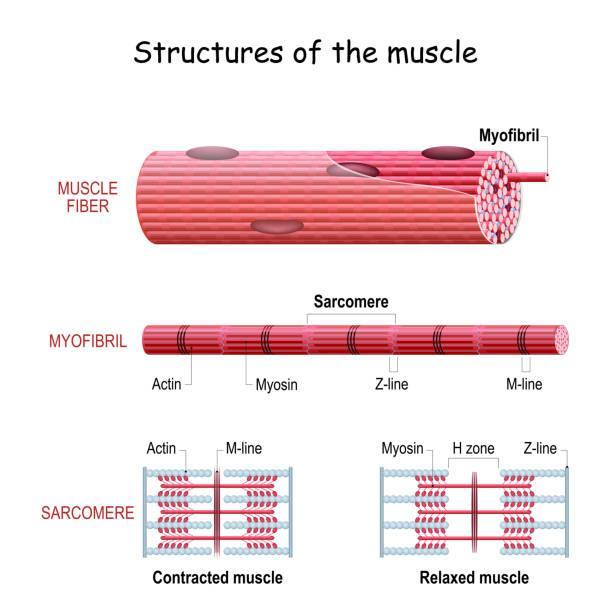
Understanding Muscle Structure and the Sarcomere
At the heart of muscle contraction lies the sarcomere—the basic contractile unit in muscle fibres. Each sarcomere is made up of overlapping thin (actin) and thick (myosin) filaments arranged in bands:
A-band: The central region where thick and thin filaments overlap and remain constant in length during contraction.
I-band: Contains only thin filaments and shortens during contraction.
H-zone: A lighter area in the middle of the A-band, containing only thick filaments; it narrows as contraction occurs.
Z-lines: The boundaries that hold the sarcomere together, giving muscles their striped appearance.
M-line: Located in the centre of the sarcomere, providing structural support by holding the thick filaments together.
These structures not only define the sliding filament theory diagram but also explain how muscle length changes when the filaments slide past one another.
Read More: Mitosis and Meiosis
The Sliding Filament Theory Explained Simply
In simple terms, muscle contraction occurs when the actin (thin) filaments slide over the myosin (thick) filaments, shortening the sarcomere. This process does not involve the filaments themselves expanding or contracting; rather, it is the relative movement between them that produces the force for contraction.
For Class 11 students, understanding that this theory is a cornerstone of muscle physiology is essential. Our explanation of the sliding filament theory's simple mechanism will help you grasp the key principles without unnecessary complications.
Explore Difference Between Actin and Myosin
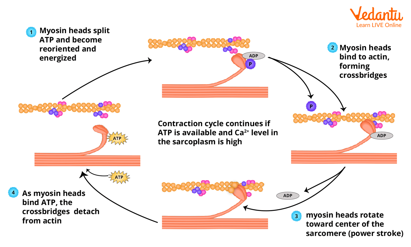
The Detailed Sliding Filament Theory Steps
Signal Initiation at the Neuromuscular Junction: A nerve impulse arrives at the neuromuscular junction, releasing neurotransmitters that trigger an electrical impulse in the muscle fibre.
Calcium Ion Release: The impulse causes the sarcoplasmic reticulum to release calcium ions, which bind to troponin on the actin filaments. This binding shifts tropomyosin, exposing the myosin-binding sites.
Cross-Bridge Formation: Energised myosin heads attach to these newly exposed sites on the actin, forming cross-bridges—a critical phase in the sliding filament theory steps.
Power Stroke and Filament Sliding: The myosin heads pivot, pulling the actin filaments towards the centre of the sarcomere. This action is powered by ATP hydrolysis and is the primary motion described by the sliding filament theory steps.
Detachment and Re-cocking: A new ATP molecule binds to the myosin head, causing it to detach from the actin filament. The myosin head then re-cocks, ready to form another cross-bridge, continuing the cycle until muscle contraction is complete.
Each of these sliding filament theory steps is vital for producing smooth, coordinated muscle movement.
The Historical Perspective: Sliding Filament Theory Proposed by Pioneers
The sliding filament theory was first proposed by researchers in the 1950s—most notably by Andrew Huxley, Hugh Huxley, and Rolf Niedergerke. These pioneers demonstrated how the overlapping of actin and myosin filaments results in the shortening of muscle fibres during contraction. Today, when we refer to the sliding filament theory proposed by these experts, we acknowledge a significant milestone in our understanding of muscle physiology.
Over time, further research has refined our knowledge of the sliding filament theory proposed by these early scientists, reinforcing its status as a fundamental concept in biology.
The Role of ATP, Myosin, and the Neuromuscular Junction in Muscle Contraction
A key element in muscle contraction is ATP—the molecule that fuels the action of myosin. Myosin, a motor protein, converts the chemical energy from ATP into mechanical work. During contraction, ATP binds to the myosin head, allowing it to detach from actin and then reattach to form a new cross-bridge. This cycle repeats, moving.
The neuromuscular junction plays an equally critical role. It is the site where nerve impulses initiate the contraction process. Understanding the interactions at the neuromuscular junction is crucial for appreciating how signals translate into the sliding filament theory physiology of muscle movement.
Additional Insights into Sliding Filament Theory Physiology
Beyond the basic mechanics, the sliding filament theory physiology encompasses several regulatory mechanisms:
Calcium Regulation: Calcium ions not only enable cross-bridge formation by binding to troponin but also regulate the speed and strength of contraction.
Energy Efficiency: The continuous cycle of ATP hydrolysis and cross-bridge cycling ensures that muscles can respond quickly to stimuli.
Structural Proteins: Proteins such as titin and nebulin help maintain the structural integrity of the sarcomere and regulate filament alignment during contraction.
Unique Aspects and Clinical Relevance
While many texts cover the basics, here are some unique insights that set this guide apart:
Clinical Connections: Abnormalities in the muscle contraction process can lead to conditions such as myopathies and muscular dystrophies. Understanding the sliding filament theory physiology helps in appreciating how these conditions affect movement.
Exercise Physiology: The efficiency of the cross-bridge cycle underlies improvements in muscle strength and endurance. Thus, the sliding filament theory's simple explanation is also crucial for sports science.
Advanced Research: Recent studies have expanded on the sliding filament theory proposed by early researchers, exploring how variations in filament overlap can affect the force produced by a muscle.
Related Links:


FAQs on Sliding Filament Theory in Muscle Contraction
1. What is the sliding filament theory?
The sliding filament theory explains that muscle contraction occurs when thin actin filaments slide past thick myosin filaments, shortening the muscle fiber without changing filament length. This theory describes how skeletal muscles contract at the molecular level.
- Actin and myosin overlap increases during contraction.
- The sarcomere shortens as Z-lines move closer together.
- The length of actin and myosin filaments remains constant.
2. How does the sliding filament theory work step by step?
The sliding filament theory works through a cyclic interaction between actin and myosin called the cross-bridge cycle. The steps include:
- Calcium release from the sarcoplasmic reticulum binds to troponin.
- Tropomyosin shifts, exposing binding sites on actin.
- Myosin heads bind to actin forming cross-bridges.
- Using ATP, myosin pulls actin inward (power stroke).
- A new ATP molecule causes detachment and re-cocking of the myosin head.
3. What is the role of actin and myosin in the sliding filament theory?
In the sliding filament theory, actin forms the thin filament and myosin forms the thick filament responsible for force generation. Their roles include:
- Actin provides binding sites for myosin heads.
- Myosin heads attach to actin and perform the power stroke.
- ATP binding and hydrolysis by myosin drive filament sliding.
4. What happens to the sarcomere during muscle contraction?
During muscle contraction, the sarcomere shortens as actin filaments slide toward the center of the M-line. The changes include:
- Z-lines move closer together.
- The I-band becomes shorter.
- The H-zone may disappear.
- The A-band remains the same length.
5. Why is ATP important in the sliding filament theory?
ATP is essential in the sliding filament theory because it provides energy for myosin movement and cross-bridge cycling. Specifically:
- ATP binding causes myosin to detach from actin.
- ATP hydrolysis re-cocks the myosin head.
- Energy released drives the power stroke.
6. What is the role of calcium ions in muscle contraction?
Calcium ions trigger muscle contraction by exposing binding sites on actin filaments. When released from the sarcoplasmic reticulum:
- Calcium binds to troponin.
- This causes tropomyosin to shift position.
- Myosin can then bind to actin.
7. What is the cross-bridge cycle in the sliding filament theory?
The cross-bridge cycle is the repeating interaction between myosin heads and actin that produces muscle contraction. It involves:
- Attachment of myosin to actin.
- The power stroke that pulls actin inward.
- Detachment after ATP binds.
- Reactivation of the myosin head through ATP hydrolysis.
8. What is the difference between the A-band, I-band, and H-zone?
The A-band, I-band, and H-zone are regions of a sarcomere that change differently during contraction. Their differences are:
- A-band: Length of the myosin filament; remains constant.
- I-band: Region with only actin; shortens during contraction.
- H-zone: Central region with only myosin; decreases or disappears during contraction.
9. Does the length of actin and myosin filaments change during contraction?
No, the length of actin and myosin filaments does not change during muscle contraction. Instead:
- Filaments slide past each other.
- Their overlap increases.
- The sarcomere shortens without filament shortening.
10. In which types of muscles does the sliding filament theory apply?
The sliding filament theory applies to skeletal muscle, cardiac muscle, and, with modifications, smooth muscle. Specifically:
- Skeletal and cardiac muscles have organized sarcomeres.
- Smooth muscle lacks visible sarcomeres but still uses actin–myosin interactions.
- All three types rely on ATP and calcium for contraction.


