


What is the Cornea in the Eye and Why is it So Important for Vision?
The cornea in the human eye is the clear, dome-shaped front covering of the eyeball. It acts like a transparent protective shield and also plays a major role in focusing light so that vision remains sharp and clear. Because it is positioned at the very front of the eye, the cornea is the first structure that incoming light passes through before reaching deeper parts such as the aqueous humor, iris, pupil, lens, and finally the retina.
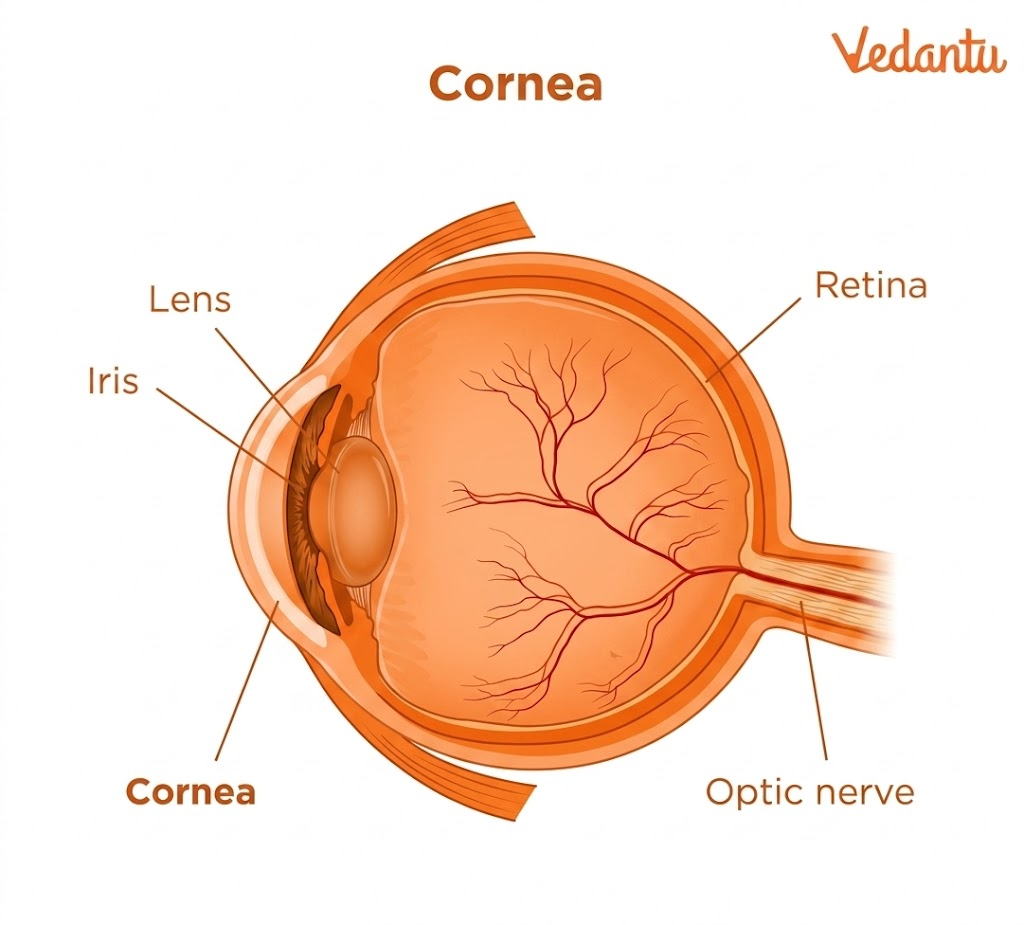
Also Read: Structure of the Human Eye
Function of Cornea
The function of cornea is one of the most important parts of this topic. The cornea has several major roles in vision and protection.
1. Helps Focus Light
The most important function of the cornea is to bend or refract incoming light. This refraction is essential for focusing the light correctly onto the retina. Without the cornea, the eye would not be able to form a properly focused image.
2. Protects the Inner Eye
The cornea acts as a clear protective barrier. It allows light to pass, but blocks many harmful particles such as dust, debris, and germs. This is why it is often compared to a windshield.
3. Filters Some UV Rays
The cornea also filters some ultraviolet radiation, helping protect internal eye tissues from potential damage.
4. Supports the Eye’s Optical Power
Its dome shape is not accidental. That curve contributes strongly to the optical performance of the eye and is essential for sharp vision.
5. Triggers Protective Reflexes
Because the cornea is extremely sensitive, it quickly detects pain or irritation. This allows the body to react rapidly through blinking, tearing, and avoidance responses.
Structure of Cornea
The structure of the cornea is remarkable because it is both strong and transparent. It must maintain its shape, stay clear for vision, and also protect the eye surface. This is made possible by its multiple-layered arrangement.
The cornea has six layers, and each one has a specific role. Together, these layers work like laminated safety glass, where multiple thin layers combine to create strength and functional stability.
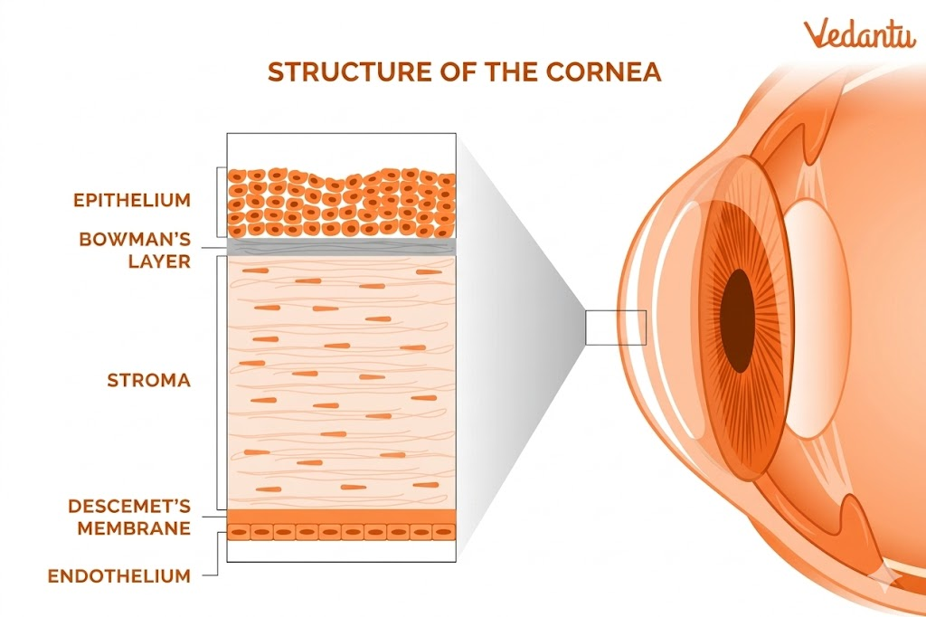
6 Layers of Cornea
The 6 layers of cornea are:
Epithelium
Bowman’s layer
Stroma
Pre-Descemet’s layer
Descemet’s layer
Endothelium
This six-layer model is important because older descriptions often focused on fewer layers. The inclusion of the pre-Descemet’s layer, also called Dua’s layer, reflects a more recent understanding of corneal anatomy.
Detailed Functions of Each Corneal Layer
1. Epithelium
The epithelium is the outermost layer of the cornea. It acts as the first physical barrier between the eye and the outside world. It is extremely sensitive to pain, which is protective because even a minor irritation can trigger a quick response such as blinking or tearing. Researchers estimate that the cornea has about 300 to 600 times as many pain receptors as skin, showing how sensitive this layer is.
2. Bowman’s Layer
Bowman’s layer is a tough, collagen-rich layer that helps provide structural support. It contributes to maintaining corneal shape and adds strength to the corneal surface.
3. Stroma
The stroma is the thickest layer of the cornea. It is extremely important because it provides much of the cornea’s structural strength and also helps refract light. Since it forms the main bulk of the cornea, it is central to both optical performance and mechanical stability.
4. Pre-Descemet’s Layer
The pre-Descemet’s layer or Dua’s layer is a strong barrier separating the internal eye environment from the outside. Research suggests that it is airtight and structurally distinct. Although some experts still debate whether it should be considered an independent layer or part of the stroma, surgeons now take it into account during corneal surgery planning.
5. Descemet’s Layer
Descemet’s layer is thin, elastic, and strong. It supports eye structure and helps protect the inner parts of the eye from injury and infection.
6. Endothelium
The endothelium plays a key role in fluid balance. It helps regulate the amount of water and fluid in the stroma so that the cornea remains clear and functions properly. If fluid balance is disturbed, corneal transparency can be affected.
Why is the Cornea So Sensitive?
One of the most distinctive features of the cornea is its high sensitivity. This sensitivity is mainly protective. Since the cornea is the first exposed surface of the eye, it must respond very quickly to harmful stimuli such as dust, foreign particles, sharp objects, heat, chemicals, or UV damage. That is why the outer corneal layer contains a very high density of pain receptors.
This sensitivity helps trigger immediate protective actions such as:
blinking
tearing
eye closure
turning away from the irritant
In biological terms, this sensitivity is an important survival feature.
Common Corneal Disorders
The cornea is vulnerable to a wide range of injuries, infections, inflammatory conditions, and structural diseases.
1. Dry Eye
The epithelium depends on tear fluid for lubrication and oxygen absorption. When the eye surface becomes too dry, the cornea becomes painful and vision may be disturbed.
2. Infections
If the corneal surface is damaged, germs such as bacteria, viruses, fungi, and parasites can infect it. One example mentioned is acanthamoeba keratitis, a parasitic infection.
3. Keratitis
Keratitis means inflammation of the cornea. It may result from infection, trauma, or other disease conditions.
4. Injuries
The cornea may develop:
abrasions or scratches
lacerations
ulcers
erosions
5. Environmental Damage
The cornea can be damaged by:
extreme heat
extreme cold
ultraviolet exposure
chemicals in gas or liquid form
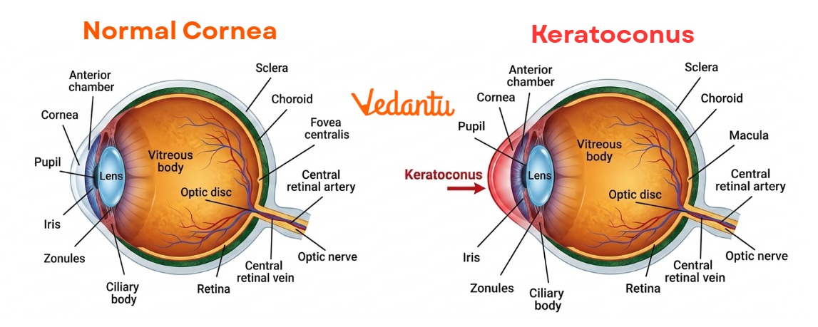
6. Corneal Dystrophies
These are structural disorders that affect how the cornea is built or how its layers interact. There are more than 20 such diseases, including keratoconus and Fuchs’ dystrophy.
Treatments for Corneal Conditions
Treatment depends on the specific condition, but common options include:
Over-the-Counter Remedies
Simple irritation or dry eyes may be managed with basic supportive products.
Medications
Infections and some inflammatory conditions may be treated with:
eye drops
ointments
oral medicines
Protective or Wearable Treatments
These include:
eye patch
scleral lenses
bandage contact lenses
amniotic membrane grafts
1. Laser Surgery
Procedures such as LASIK reshape the cornea to change how it refracts light.
2. Eye Surgery
Surgery may repair damage or correct disease.
3. Cornea Transplant or Artificial Cornea
In severe cases, if the cornea becomes cloudy or loses proper refractive function, replacement surgery may be needed.
How to Protect the Cornea?
Corneal protection is a major part of eye health.
1. Wear Protective Eyewear
Safety glasses or goggles are strongly recommended when:
using tools or machines
doing yardwork
handling chemicals
working with heat or open flames
using compressed air or water
playing sports
facing UV exposure such as reflected sunlight from water or snow
2. Follow Basic Safety Habits
read instructions before using chemicals or machines
use the right type of eye protection
wash hands frequently
do not drive if an eye injury may affect vision
seek medical care for burns near the eye
3. Keep Corneas Healthy
get regular eye exams
wash hands often
do not share cosmetics
wear and store contact lenses properly
avoid rubbing or touching the eyes
Symptoms That Need Medical Attention
A person should seek medical attention if any of the following occur:
blurred, double, clouded, or distorted vision
sudden vision loss
foreign body sensation
eye pain
watery eyes
light sensitivity
eye redness or irritation
visible bleeding or fluid in abnormal eye spaces
getting hit directly on or around the eye
visible puncture, wound, or tear on the eye surface
These signs may indicate corneal injury or disease and should not be ignored.
What to Do If Something Gets Stuck in the Eye?
If something feels stuck in the eye:
Do:
blink slowly and deliberately
flush with saline or clean water if there is no visible injury
use a cup as a protective cover while seeking help if needed
Do Not:
rub the eye
touch the eyeball directly
use a spray nozzle directly on the eye
rinse or attempt removal if there is a visible puncture or wound
These simple steps can reduce further damage before proper care is received.
FAQs on Cornea in Human Eye: Functions, 6 Layers, Diagram and Common Disorders
1. What is the function of the cornea?
The cornea helps focus light by bending (refracting) it as it enters the eye. It also protects the eye and keeps the surface moist with the help of tear fluid.
2. Can we see without a cornea?
No, we cannot see properly without the cornea because it is essential for focusing light and protecting the inner parts of the eye.
3. Can you fix a damaged cornea?
Yes, a damaged cornea can often be treated. Minor injuries heal naturally, while serious damage may require medicines, laser treatment, or a cornea transplant.
4. What happens if your cornea is damaged?
Damage to the cornea can cause pain, redness, sensitivity to light, and blurred vision. Severe damage may lead to infections, scarring, or vision loss.
5. What are the symptoms of a bad cornea?
Common symptoms include blurry vision, eye pain, redness, watery eyes, light sensitivity, and a feeling that something is stuck in the eye.
6. What is the best treatment for corneal erosion?
Treatment usually includes lubricating or salt-based eye ointments, medicines like antibiotics or steroids, and sometimes special contact lenses to help healing.
7. What are the 4 stages of a corneal ulcer?
The four stages are:
Surface damage (epithelial defect)
Infection spreading deeper (stromal infiltration)
Severe thinning (descemetocele)
Full perforation of the cornea
8. What are the first signs of a torn retina?
Early signs include sudden flashes of light, floating spots in vision, reduced vision, and loss of side (peripheral) vision.
10. How can I avoid mixing up cornea and sclera under exam pressure?
To avoid confusion:
• Remember that the cornea is the transparent, curved front window of the eye.
• The sclera is the white, opaque outer layer surrounding most of the eyeball.
• Visualize the cornea as the clear, focusing surface and the sclera as the protective white covering.
Mnemonics or labelled diagrams aid quick recall.